
The Trust is indebted to Dr Catherine Guly, Consultant Ophthalmic Physician, Bristol Eye Hospital for providing the following information on vasculitis and the eye
Vasculitis can affect different parts of the eye. In some patients there is only mild inflammation which does not affect the vision and in others symptoms are more severe and the vision may become affected. With advances in the understanding of ocular inflammation it has become clear that the treatments for vasculitis elsewhere in the body are also useful for treating vasculitis affecting the eye. This article discusses the different forms of vasculitis and explains how they can affect the eye.
How is vasculitis of the eye diagnosed?
Vasculitis affecting the eye is usually diagnosed by an ophthalmologist (eye doctor). Ophthalmologists use a slit lamp which has a microscope that gives a magnified view of the eye. It is possible to look into the back of the eye by enlarging the pupil with dilating eye drops.
The eye examination can tell which part of the eye is inflamed but does not show what has caused the inflammation and so other investigations, such as blood tests, can be helpful in making a diagnosis. Blood markers of inflammation (including the CRP, ESR and plasma viscosity) are useful in diagnosing and monitoring giant cell arteritis. If there is inflammation behind the eye a CT or MRI scan can be useful. Rarely, a small sample of tissue is taken from the eye or around the eye to send for analysis in the laboratory to look for signs of vasculitis. In Giant cell arteritis a sample of artery from the temple (the temporal artery) is used to help with the diagnosis.
How do different forms of vasculitis affect the eye?
Giant cell arteritis (temporal arteritis) can result in an optic neuropathy in one or both eyes. An optic neuropathy is a disruption of the function of the optic nerve, in this case due to inflammation of the arteries that supply blood to the optic nerve. The optic nerve joins the eye to the brain.
Patients with an optic neuropathy due to giant cell arteritis may notice loss of vision or transient loss of vision in one or both eyes. This is often associated with a headache. There may be pain on eating if the blood supply to the jaw is also affected. Early treatment with steroids usually stabilises the vision but if there is damage to the optic nerve it does not always recover. Occasionally, giant cell arteritis disrupts the blood supply to the retina (retinal artery occlusion) or results in double vision (cranial nerve palsy).
ANCA associated vasculitis (Wegener’s granulomatosis and Microscopic polyangitis) and Polyarteritis nodosa can affect all the different parts of the eye. The most common symptoms of vasculitis are redness and eye pain. There may be increased sensitivity to light.
Some forms of inflammation are mild and do not affect the vision. For example, episcleritis is the inflammation of the outside coat of the eye, the episclera, and may result in a red, irritated eye. Other forms of inflammation are more serious and may affect the vision if untreated. These include inflammation of the cornea which is the clear window at the front of the eye (keratitis), inflammation inside of the eye (uveitis) or inflammation of the sclera which is the white outer coat of the eye (scleritis). Scleritis in particular may cause severe pain around the eye, although this usually settles with treatment.
Orbital inflammation is inflammation in the eye socket. With orbital inflammation there may be double vision, the eyelid may be higher or lower than normal and the eye may protrude more than normal. The vision can become blurred if the optic nerve is affected. Orbital inflammation is most commonly associated with Wegener’s granulomatosis.
EGPA rarely causes inflammation in the eyes.
How is vasculitis of the eye treated?
The treatment will depend on the type of vasculitis and which part of the eye is inflamed. Eye drops can be used to treat inflammation at the front of the eye such as episcleritis and some forms of uveitis. Other types of inflammation usually require high dose steroid treatment with or without other immunosuppressive medications (such as mycophenolate mofetil, cyclophosphamide or methotrexate) or biological medications (such as adalimumab, infliximab and rituximab). Giant cell arteritis can usually be treated with steroids alone but sometimes other immunosuppressive medications are added. Patients usually require high doses of treatment initially to gain control of the inflammation and then the treatment is tapered to the lowest dose that will maintain control of the inflammation.
If the patient has inflammation elsewhere in the body the ophthalmologist will aim to work closely with the other physicians involved so that as far as possible the medicines chosen will treat the inflammation in the eye as well as any inflammation elsewhere in the body. The treatment will normally be dictated by the organ which is most at threat so if the eyes are inflamed and the sight is at threat then the ophthalmologist will need to guide the type of treatment required and how quickly the treatment can be reduced.
Is there anything I can do to protect my vision?
If you have vasculitis you should report any new visual or eye symptoms to your doctor or your optometrist (optician), particularly if you notice any change in your vision. It is also a good idea to see an optometrist (optician) on a yearly basis as they will perform a thorough eye examination during your eye check. An optometrist can also detect any side effects from long term use of prednisolone (steroids), such as cataracts and more rarely, glaucoma.
More detailed medical information about how vasculitis affects different parts of the eye
All the conditions detailed here may be associated with vasculitis but are also found with other medical conditions.
Conjunctivitis is inflammation of the conjunctiva. When the conjunctiva is inflamed the eye and inside the eyelids become red and the eye may feel gritty. The vision is usually unaffected. Conjunctivitis is also very common in the general population and is usually caused by infection or allergy, but may also be caused by vasculitis.
Episcleritis is inflammation of the episclera, which is the thin covering over the sclera. With episcleritis the eye looks red, and this may affect just a patch of the episclera so that just part of the eye is red, or it may affect the whole episclera. The eye may feel irritated and uncomfortable but the vision is unaffected. Episcleritis is diagnosed with a slit lamp examination. Phenylephrine eye drops blanch the affect area (unlike scleritis where the inflammation is deeper) and this can be helpful in making a diagnosis.
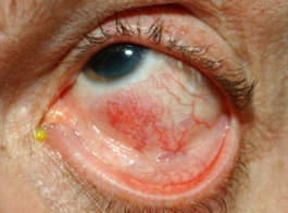
Peripheral ulcerative keratitis is inflammation and ulceration of the cornea. The eye is usually red and painful and the vision may be blurred. The inflammation and ulceration start at the edge of the cornea and in severe cases the cornea may perforate. Keratitis is diagnosed with a slit lamp examination. Corneal ulcers are detected using fluorescein eye drops; the epithelial defect takes up the stain and the ulcer glows yellow under a blue light.
Scleritis is inflammation of the sclera. When the sclera becomes inflamed the eye usually becomes red and painful. The pain often disturbs sleep and sometimes the vision becomes blurred. Occasionally, only the back part of the sclera becomes inflamed and this is called posterior scleritis. The posterior sclera is not visible from the outside of the eye and in patients with posterior scleritis the eye may be painful but does not look red. Scleritis is diagnosed using a slit lamp examination. An ultrasound scan of the eye is useful to detect posterior scleritis.
Uveitis is inflammation inside the eye. The ‘uvea’ is the pigmented layer inside the eye made of the choroid, ciliary body and iris, but uveitis also affects other parts of the eye like the retina and so it is easier to think of it as ‘intraocular inflammation’. Uveitis can be divided up into anterior uveitis which affects the front chamber of the eye, intermediate uveitis which affects the middle part of the eye (the vitreous) and posterior uveitis where the back of the eye (retina and/ or choroid) is inflamed. In panuveitis the inflammation affects the front, the middle and the back of the eye.
Uveitis usually causes a red painful eye which is worse in bright light but in some forms of uveitis the eyes are white and there is no pain. If there is inflammation in the vitreous there may be multiple floaters that obscure the vision and if there is inflammation in the retina or choroid the vision may be blurred or there may be loss of vision.
The diagnosis is made using a slit lamp examination, and additional imaging of the retina and choroid (using fluorescein angiography, indocyanine green angiography and optical coherence tomography imaging) is helpful in some cases.
Retinal artery occlusion is where there is a disruption of the blood supply to the eye. This is rare in vasculitis but it results in sudden loss of vision. Further investigations are usually required to determine if the occlusion is primarily due to vasculitis or if there may have been an embolus (clot travelling to the artery from elsewhere in the body) resulting in the occlusion.
Optic neuropathy is an abnormality of the function of the optic nerve. Patients with optic neuropathy usually notice loss of vision, which can be sudden. Sometimes patients have transient vision loss. Patients may be aware of blurred or patchy vision and may notice that colours seem washed out. An optic neuropathy is diagnosed through tests of optic nerve function, including visual acuity, colour vision, visual fields and pupil examination. The optic nerve may appear swollen or pale. Other investigations which may be helpful include electrophysiology (to measure optic nerve function), MRI/ CT scan of the head and orbits and a lumbar puncture.
Orbital inflammation is where there is inflammation in the eye socket, which includes the muscles around the eye. With orbital inflammation there may be double vision, the eyelid may be higher or lower than normal and the eye may protrude more than normal. There may be pain or discomfort around the eye and if there is any pressure on the optic nerve there may be an associated optic neuropathy. The eyelids and conjunctiva may also be inflamed.
Cranial nerve palsies. Vasculitis can disrupt the blood supply to nerves around the eye which supply the muscles that move the eye (the third nerve, the fourth nerve and the sixth cranial nerves). Damage to any of these nerves usually results in double vision. If the third nerve is affected there may also be a droopy eyelid (ptosis) and enlarged pupil on the affected side.